banner img

X-Ray Department

  • Home
  • X-Ray Department

The X-ray Department is a vital part of a healthcare facility that uses radiology to help diagnose and monitor various medical conditions. X-ray imaging is one of the most common and widely used diagnostic tools, providing clear and detailed images of the internal structures of the body, especially bones, tissues, and organs. The X-ray department plays a key role in patient care by providing essential images for medical evaluation, treatment planning, and monitoring of diseases or injuries.

Key Functions of the X-ray Department

  1. Diagnostic Imaging The X-ray department’s primary role is to provide diagnostic images using X-ray technology. X-ray is especially effective in detecting:

    • Bone fractures: Fractures and breaks in bones are commonly diagnosed using X-ray imaging.
    • Joint issues: X-rays are used to assess joint injuries, dislocations, or degenerative conditions like arthritis.
    • Lung conditions: Chest X-rays are commonly used to identify conditions like pneumonia, tuberculosis, or lung cancer.
    • Infections and tumors: Certain X-ray techniques can detect tumors, infections, or other abnormalities in organs and tissues.
    • Dental issues: X-rays are also used in dental offices to examine teeth, gums, and jawbones for cavities, infections, and other dental concerns.
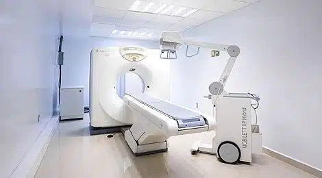
  2. X-ray Procedures Various types of X-ray procedures are performed in the X-ray department, depending on the area of the body that needs to be examined. These include:

    • Plain X-ray (Conventional X-ray): The most basic type, used to capture images of bones and internal organs.
    • Fluoroscopy: A real-time X-ray imaging technique that allows continuous imaging of a body part in motion, often used for guiding procedures or examining the gastrointestinal system.
    • Mammography: A specialized type of X-ray used to examine breast tissue, often for screening for breast cancer.
    • Computed Tomography (CT) Scan: A more advanced X-ray procedure that combines multiple X-ray images taken from different angles to create cross-sectional images of the body. It is commonly used for examining soft tissues and organs.
    • Interventional Radiology: Involves using X-ray to guide procedures like biopsies, drainage, or catheter placements.
  3. Preparing Patients

    • Patient Assessment: Before the X-ray procedure, patients are assessed by the X-ray technician or radiographer to determine the type of imaging required.
    • Instructions: Patients may be asked to remove certain clothing or wear a hospital gown to prevent interference with the imaging. In some cases, patients may need to change their position or hold their breath during the X-ray to ensure high-quality images.
    • Safety Considerations: Pregnant women or those who suspect they may be pregnant must inform the medical team, as X-rays can harm developing fetuses. In certain situations, alternative imaging methods such as ultrasound or MRI may be recommended.
  4. Radiation Safety The X-ray department takes extensive precautions to ensure the safety of both patients and staff from radiation exposure:

    • Lead Shields: Lead aprons or shields are used to protect areas of the body that are not being imaged.
    • Radiation Dose Control: Modern X-ray machines are designed to use the lowest possible radiation dose to obtain clear images, minimizing patient exposure.
    • Staff Protection: X-ray technologists and radiologists work in designated areas, and use protective gear like lead aprons and gloves when necessary, to limit their exposure to radiation.
  5. Interpreting X-ray Images After the images are captured, they are analyzed and interpreted by a radiologist, a medical professional trained in interpreting diagnostic images. The radiologist assesses the X-ray images for signs of:

    • Fractures or dislocations
    • Abnormal growths, such as tumors
    • Infections or inflammation
    • Internal bleeding
    • Bone density issues or osteoporosis
    • Pulmonary conditions, such as pneumonia or lung infections
  6. Collaboration with Other Departments The X-ray department frequently collaborates with other departments within the healthcare facility:

    • Orthopedics: X-rays are often used by orthopedic surgeons to assess fractures, joint injuries, and other musculoskeletal issues.
    • Emergency Room: The ER department commonly utilizes X-ray imaging to quickly diagnose traumatic injuries like fractures or internal bleeding.
    • Cardiology: Chest X-rays are used to evaluate heart and lung conditions, such as heart failure or pulmonary edema.
    • Oncology: X-rays can help detect tumors or metastases in various organs, assisting oncologists in cancer diagnosis and treatment planning.
  7. Digital X-ray Systems The shift from traditional film-based X-ray systems to digital X-ray technology has greatly improved the efficiency and quality of imaging:

    • Digital Radiography (DR): Uses digital sensors to capture X-ray images, providing quicker results, higher resolution, and easier storage and sharing of images.
    • Picture Archiving and Communication System (PACS): A digital system that allows medical images to be stored and accessed electronically, enabling faster diagnosis and remote consultation between healthcare providers.
  8. Record Keeping and Reporting X-ray departments are responsible for maintaining accurate records of imaging procedures, results, and reports. These records must be securely stored and easily accessible for future reference, follow-up treatments, or legal purposes.

Roles in the X-ray Department

  1. Radiologic Technologists (Radiographers) Radiologic technologists are responsible for operating X-ray machines, positioning patients for imaging, and ensuring that high-quality images are captured. They also provide information to patients and assist with any necessary preparations before the procedure.

  2. Radiologists Radiologists are medical doctors who specialize in interpreting X-ray images and other diagnostic imaging techniques. They provide detailed reports based on the images and collaborate with other healthcare providers to make diagnoses and guide treatment plans.

  3. Medical Assistants In some cases, medical assistants may help with patient preparation and assist technologists in performing basic tasks like taking medical histories or managing equipment.

  4. X-ray Department Supervisor/Manager The supervisor or manager oversees the day-to-day operations of the X-ray department, ensuring that the team is efficient, safe, and compliant with regulatory standards. They may also be involved in training staff and managing schedules.

Conclusion

The X-ray department is an essential component of modern healthcare, offering crucial diagnostic information to support accurate diagnoses and effective treatment plans. By using advanced imaging techniques, such as conventional X-rays, CT scans, and mammography, the department plays a critical role in detecting fractures, diseases, infections, and tumors. Through collaboration with other medical professionals, attention to safety, and ongoing advancements in technology, the X-ray department contributes significantly to patient care and clinical decision-making.

Ask Any Question If You Have

Your email address will not be published. Required fields are marked *